A noninvasive MRI technique that measures volumetric changes in paraspinal muscles could transform how doctors assess nerve damage in the cervical spine, which helps guide more precise treatment decisions and potentially speed recovery for patients suffering from either sudden trauma or gradual degeneration.
Cervical radiculopathy, whether from degenerative spinal disease or traumatic brachial plexus injury caused by traffic accidents, often limits movement in both young adults and seniors. The condition can severely affect quality of life and place economic and emotional strain on patients as well as their families. Clinically, determining whether a nerve root is normal, partially or completely avulsed by nonquantified radiological evaluation has been challenging, often forcing surgeons to revise plans during surgery.
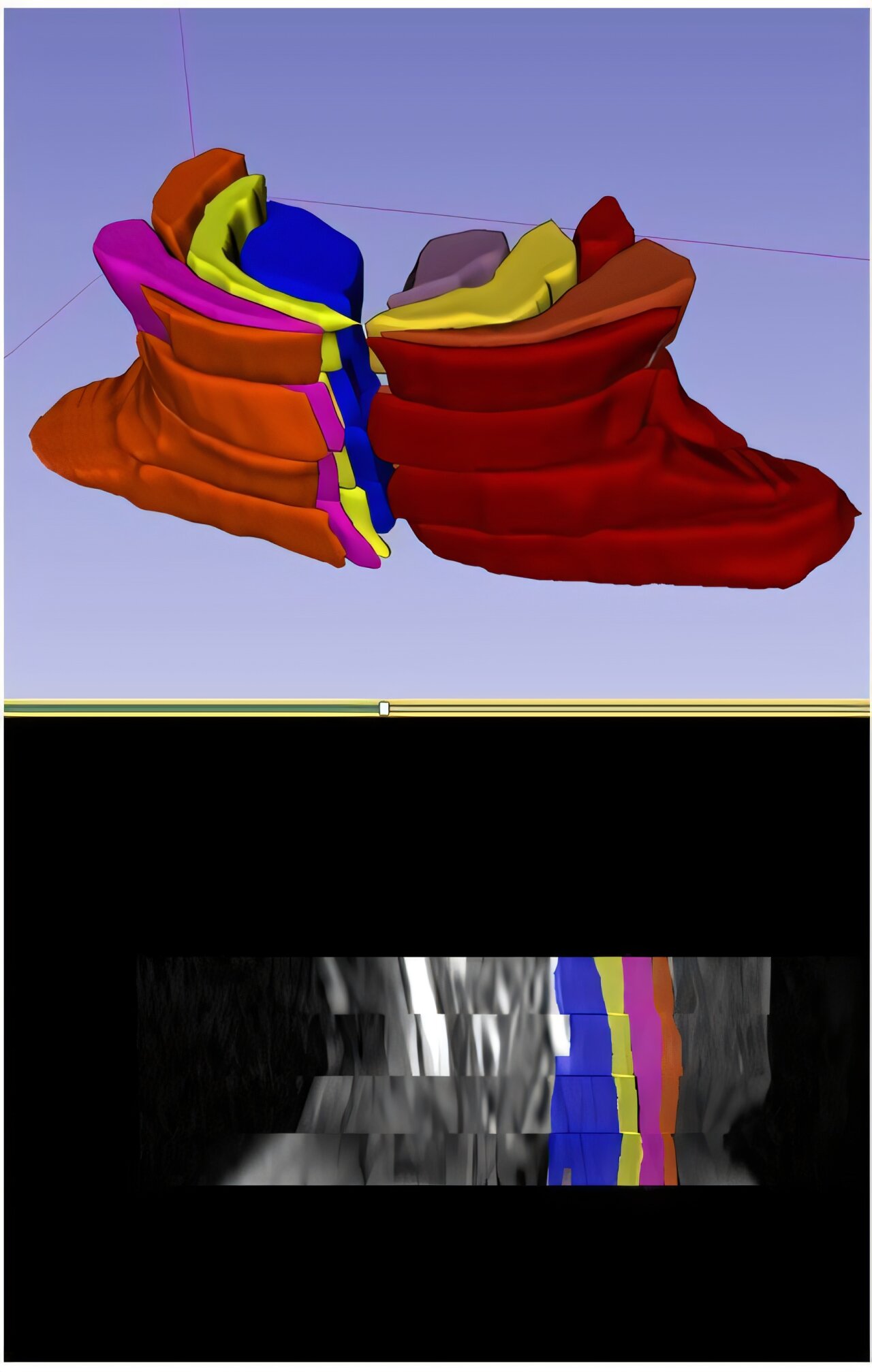
A multidisciplinary research team at Chang Gung Memorial Hospital and National Taiwan University has now refined a quantitative add-on to standard MRI exams through a noninvasive analysis of deep paraspinal muscles adjacent to the cervical spine. By comparing muscle volumes between the affected and healthy sides, they established precise numerical cutoffs: a ratio of 0.95 distinguishes intact from damaged nerves, while 0.80 differentiates partial from total avulsions. These volumetric ratios provide an accurate imaging biomarker to confirm the nerve root integrity before surgery. The study is published in Radiology.
In a retrospective study of 145 adults (mostly men aged 18–72) with upper-limb nerve trauma, the technique analyzed muscles at four cervical levels (C4–C7). It identified that the deepest muscle layer, including the semispinalis cervicis and multifidus, where muscle volume decreased consistently with nerve injury severity. Statistical analysis confirmed significant differences between intact, partial, and total avulsions (P < .001 for C4–C7).
Compared with traditional neurological or physical exams (AUC 0.80 vs. 0.59) and standard MRI reports (AUC 0.85 vs. 0.76), the innovative method achieved AUC values of 0.88 and 0.91, correctly classifying all four cervical levels in 55% of cases and three levels in 29%, which closely matches surgical findings and improving preoperative planning.
Beyond trauma, the approach may extend to degenerative conditions such as disk herniation or spinal stenosis. The same muscle-volume ratios could reveal early atrophy from chronic compression, helping clinicians decide between conservative management or early intervention. In aging populations like Taiwan's, where spinal disorders are increasing, this cost-effective technique could integrate seamlessly into routine MRI scans.
Looking ahead, the team plans to incorporate artificial intelligence to automate segmentation and ratio calculations, potentially reducing analysis time from hours to minutes while maintaining diagnostic precision.
"This new assessment tool, developed through multidisciplinary collaboration, opens a window for noninvasive and quantitative evaluation of nerve root conditions," said Prof. Hsiang-Kuang Tony Liang, corresponding author of the study.
To see article on Medical Xpress: https://medicalxpress.com/news/2025-10-mri-based-accurately-quantifies-paraspinal.html